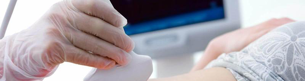
Ultrasound, also known as sonography, is a widely used medical imaging technique that has several applications in the field of pathology. Pathology is the study of diseases and abnormalities in the body, and ultrasound can be a valuable tool for diagnosing and monitoring various conditions. Here are some of the ways ultrasound is used in pathology:
- Diagnostic Imaging: Ultrasound is commonly used to visualize and assess various organs and tissues in the body. It can help pathologists identify abnormalities such as tumors, cysts, and other structural issues. For example, it is frequently used in the evaluation of the liver, gallbladder, kidneys, thyroid, and reproductive organs.
- Guidance for Biopsies: Ultrasound can be used to guide the placement of a biopsy needle when a tissue sample needs to be collected for pathological analysis. This is particularly useful in cases where a suspicious mass or lesion is identified, and a pathologist needs a tissue sample for a definitive diagnosis.
- Fetal Pathology: Obstetric ultrasound is used to monitor the development of the fetus during pregnancy. It can help identify congenital abnormalities and assess the overall health and growth of the fetus. Pathologists may be involved in assessing these images for any signs of fetal pathology.
- Breast Imaging: Ultrasound is often used as a complementary imaging modality alongside mammography to evaluate breast tissue. It can help in the characterization of breast masses and assist in the diagnosis of breast cancer.
- Vascular Pathology: Ultrasound can be used to assess blood vessels (vascular ultrasound) to identify conditions such as deep vein thrombosis, arterial stenosis, and aneurysms.
- Thyroid Pathology: Thyroid ultrasound is used to evaluate the thyroid gland for abnormalities, such as nodules or cysts, which can be indicative of thyroid diseases like thyroid cancer or thyroiditis.
- Musculoskeletal Pathology: Musculoskeletal ultrasound is employed to examine joints, tendons, ligaments, and muscles, helping diagnose conditions such as tendonitis, bursitis, and ligament injuries.
- Cardiac Pathology: Echocardiography, a specialized form of ultrasound, is used to assess the structure and function of the heart. It is invaluable in diagnosing heart diseases, valvular abnormalities, and cardiac defects.
- Gastrointestinal Pathology: Ultrasound can be used to investigate the gastrointestinal tract, including the assessment of the appendix for signs of appendicitis or the gallbladder for signs of cholecystitis.
- Renal Pathology: It is commonly used to evaluate kidney size and structure, detect renal cysts, and identify kidney stones and other kidney disorders.
Ultrasound is a non-invasive, radiation-free imaging technique that provides real-time images, making it a valuable tool for pathologists to diagnose and monitor a wide range of medical conditions. It is often used in conjunction with other diagnostic methods and plays a significant role in modern medical practice.